
after the operation and the villi were found to be extending into the holes in the side arms of the cannula. The following observations support the opinion that these measurements are reasonably accurate records of the oxygen tension in the duck’s intestine. tetracycline hydrochloride was given 72 hr.

500,000 units of benzyl penicillin were given by injection, post-operatively, and an injection of 250 mg.

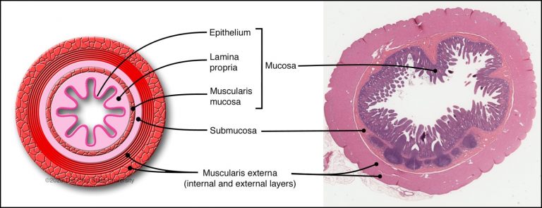
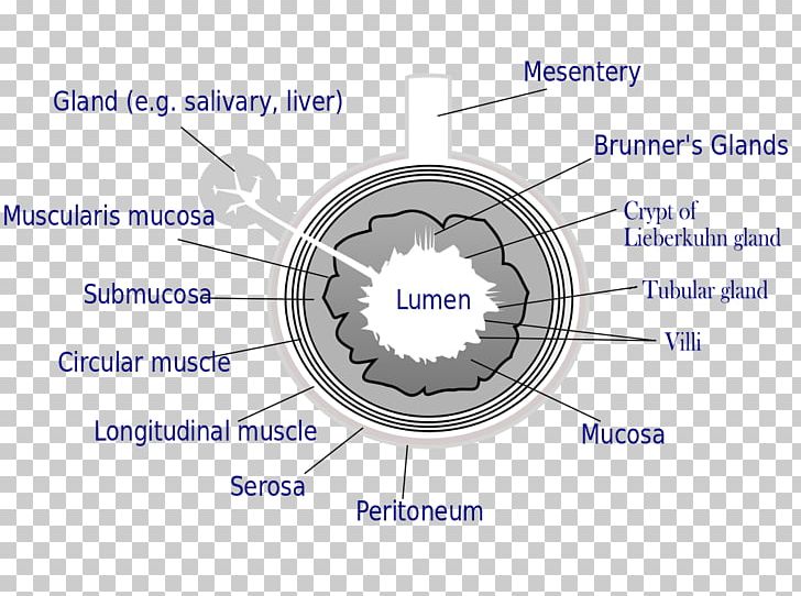
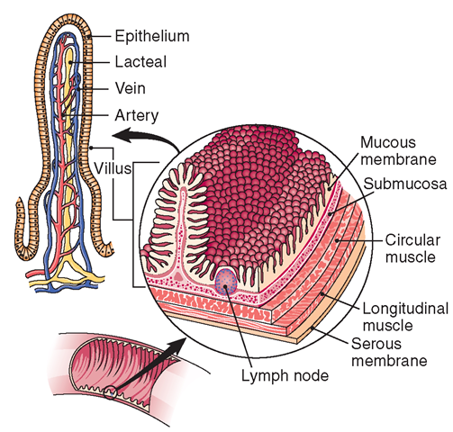
The intestine was handled as little as possible and sutures, used to close the wound in the wall of the intestine, were never positioned on the side where oxygen measurements were to be made. This procedure ensured that the cannula was situated behind the yolk stalk in the region of the intestine where the majority of P. After opening the peritoneal cavity, the cannula was placed in the fifth loop of the intestine counting the duodenal loop as the first. The general operational procedure followed that described by Beattie & Shrimpton (1958) for the preparation of caecal fistulae. MATERIALS AND METHODSĪnaesthesia was induced by an injection of thiopentone sodium (50 mg./ml./3 kg.) into the brachial vein, and was maintained, during the insertion of the cannula, by means of an ether/oxygen mixture. The method described here was devised to investigate the region where the trunk of the worm is found. thick, lies in contact with the villi on one side and with the contents of the gut lumen on the other. The remainder of the trunk of the parasite, which in nature is flattened dorso-ventrally and is about 1♵ mm. Thus, it may be postulated that a tension near this value exists in the crypts of Lieberkühn, through which the parasite burrows. The oxygen tension of venous blood of domestic ducks is about 35–40 mm. The host reacts against the proboscis by surrounding it with fibrous material, but the neck and spiny anterior portion of the trunk of the worm do not appear to stimulate a host reaction although they also are in contact with the tissues of the gut wall. They become attached to their hosts by means of a spiny proboscis which bores as far as the longitudinal muscle layer of the gut wall. The majority of the worms live in a zone consisting of about 20 % of the length of the intestine, situated posterior to the yolk stalk ( Crompton & Harrison, 1965). minutus are found in the intestinal wall and paramucosal lumen of many birds including domestic ducks. Of smooth muscle, an inner circular and outer longitudinal, forĬontinuous peristaltic activity of the small intestine.Many parasites of vertebrates live in this region of the lumen, for which Read (1950) proposed the name paramucosal lumen in order to emphasize that its physico-chemical properties are different from those of the rest of the lumen. The muscularis externa layer contains two layers The lymphatic capillariesĪre called lacteals, and absorb lipids. Products, and there is a muscularis mucosae layer Has a rich vascular and lymphatic network, which absorbs the digestive

The lamina propria which underlies the epithelium Of Lieberkuhn, which extend down to the muscularis Between the villi there are crypts, called crypts Together, these folds provide a huge surface area forĪbsorption. the lining columnar epithelial cells have fine projections on their apical surfaces called microvilli.smaller folds called villi, which are finger like mucosal projections, about 1mm long.large circular folds called plicae circulares (shown in the diagram to the right), most numerous in the upper.The pancreas and liverĪlso deliver their exocrine secretions into the duodenum. the small intestine secretes enzymes and has mucous producing glands.The small intestine is 4-6 metres long in humans. The main functions of the small intestine are digestion, absorption of food and production of gastrointestinal hormones. This is a diagram which shows the villi of the small intestine, as indicated by the arrows in the diagram above, at higher magnification.


 0 kommentar(er)
